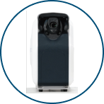
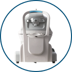
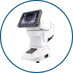
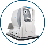
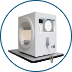
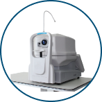
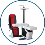
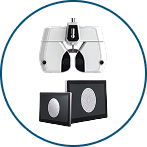
OptiWide 176 (Wide-Angle Fundus Camera)
Product Model: Reticam 3100
Top Features include
- One-Touch Operation
- Non-Mydriatic Imaging
- Fully Automated Adjustment
- Ultra-Wide HD Imaging
- True Colour Accuracy
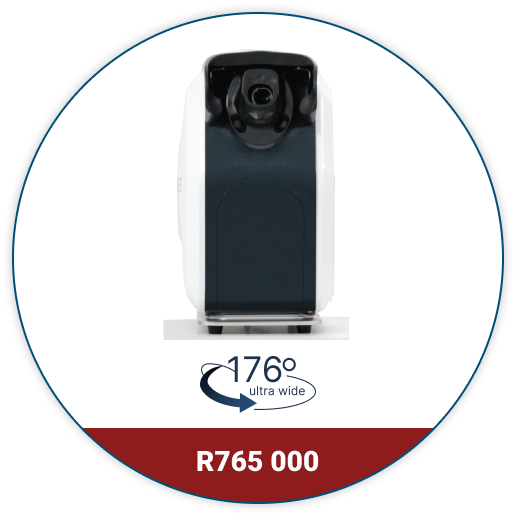
Never Seen Before
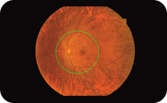
Single true colour imaging
field up to 176°
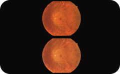
Soft Exposure to reduce pupil shrinkage after exposure
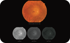
LED True Colour
& HD Resolution
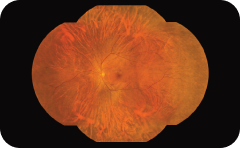
Automatic mosaic presents
a whole fundus image
High-Definition Cases, Quality First
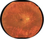
Choroidal atrophy arc on the nasal side of the optic disc in the right eye.
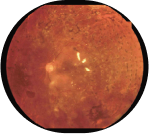
Fundus bleeding in the left eye after retinal laser photocoagulation in the left eye.
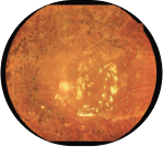
Silicone oil filling status of the left eye following retinal laser photocoagulation of the left eye.
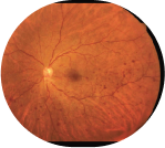
Deep retinal haemorrhage in left eye.
Specifications
| Imaging technology | LED true colour |
|---|---|
| Light source | LED white light |
| Capture mode | Single capture, automatic mosaic |
| Imaging modes | Colour image |
| Field of view (from the centre of the eyeball) | Wide field 176° (tolerance ±5%), Ultra wide field 220° (two images), Multi mosaic is larger than 260° |
| Resolution (optical) | 8 μm (tolerance ± 7%) |
| Soft exposure technology | Reduce pupil shrinkage after exposure, reduce patient discomfort, and better adapt to children and special patients |
| Working distance | 10mm ± 2mm |
| Camera flash light source illumination | The maximum illumination is less than 130000lx |
| Automatic operation | Automatic focus, Automatic alignment, Automatic capture, Automatic gain/manual |
| AI intelligent system | Accelerates precise fundus positioning, enhances focusing speed and accuracy, and reduces exam |
| Capture speed | 16 frames/second; image capture 60ms |
| Display screen | High-definition display, 27-inch colour monitor (resolution 2560*1440P) |
| Power supply | Voltage 220V, power 200W |
Note: Specifications and design are subject to change without notice.

